Proteins and Amino Acids: The Chemistry of Life's Building Blocks
Proteins do more different jobs in a living cell than almost any other class of molecule: they build muscle fiber, transport oxygen in blood, defend against infection as antibodies, and act as enzymes that drive nearly every chemical reaction your body runs. All of that functional diversity comes from combining just 20 different building blocks, called amino acids, in different sequences and lengths, the same basic strategy DNA uses with its four bases, just with a larger alphabet.
The Structure of an Amino Acid
Every amino acid shares the same basic chemical skeleton, built around a single central carbon atom (called the alpha carbon) bonded to four different groups:
- An amino group (-NH₂), which is basic and can accept a hydrogen ion.
- A carboxyl group (-COOH), which is acidic and can donate a hydrogen ion, the same functional group responsible for carboxylic acid chemistry generally.
- A hydrogen atom.
- A side chain (often called the R group), which is the only part that differs between the 20 amino acids.
The R group is what gives each amino acid its distinct chemical personality. Some R groups are nonpolar and water-repelling (like the one in valine), some are polar and water-attracting (like the one in serine), some carry a full positive or negative charge at normal biological pH (like lysine or aspartate), and some contain a ring structure (like phenylalanine). This variation in the R group is the entire source of a protein's eventual three-dimensional shape and function.
Peptide Bonds: Linking Amino Acids Into Chains
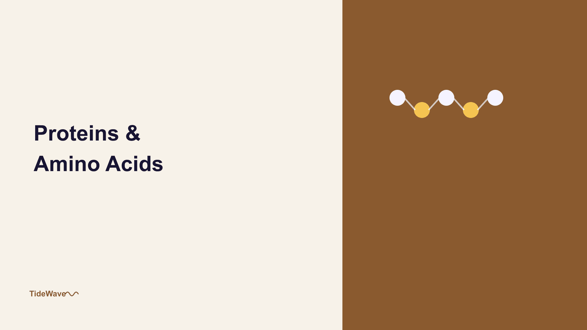
Amino acids join together through a condensation reaction (also called a dehydration synthesis), where the carboxyl group of one amino acid reacts with the amino group of another, releasing a molecule of water and forming a new covalent bond called a peptide bond:
Amino acid 1 (-COOH) + Amino acid 2 (-NH₂) → Peptide bond + H₂O
A chain of amino acids linked by peptide bonds is called a polypeptide. A functional protein is one or more polypeptide chains folded into a specific, stable three-dimensional shape.
The Four Levels of Protein Structure
Understanding a protein's structure means recognizing it operates on four distinct organizational levels, each building on the one before it.
Primary Structure
The primary structure is simply the linear sequence of amino acids in the polypeptide chain, determined directly by the sequence of a gene through the process of DNA-guided protein synthesis. Even a single amino acid substitution in this sequence can sometimes change a protein's shape enough to affect (or destroy) its function, as is the case in sickle cell disease, caused by one incorrect amino acid in a chain of nearly 150.
Secondary Structure
The secondary structure describes local, repeating folding patterns that form due to hydrogen bonding between atoms in the polypeptide backbone itself (not the R groups). Two common patterns are the alpha helix (a coiled, spring-like shape) and the beta sheet (a folded, pleated shape). These patterns form spontaneously wherever the local hydrogen-bonding geometry favors them.
Tertiary Structure
The tertiary structure is the overall three-dimensional shape of a single polypeptide chain, formed as the various R groups interact with each other and with the surrounding water. Nonpolar R groups tend to fold inward, away from water, while polar and charged R groups tend to stay on the exterior, in contact with the watery cellular environment, the same hydrophobic/hydrophilic logic that governs intermolecular forces generally. Additional stabilizing interactions include hydrogen bonds, ionic attractions between charged R groups, and occasionally strong covalent disulfide bonds between two sulfur-containing R groups.
Quaternary Structure
Some proteins are built from more than one polypeptide chain, and the quaternary structure describes how those separate chains (called subunits) arrange themselves relative to each other. Hemoglobin, the oxygen-carrying protein in blood, is a well-known example: it's made of four separate polypeptide subunits held together in a single functional unit.
Why Shape Determines Function
The recurring theme across all four structural levels is that a protein's shape determines what it can do. An enzyme's shape creates a precisely fitted pocket (called an active site) that only certain molecules can bind to. A structural protein like collagen forms long, strong fibers because of its specific, repeating helical shape. If a protein loses its correct shape, a process called denaturation, caused by heat, extreme pH, or certain chemicals, it typically loses its function entirely, even though its primary sequence of amino acids remains completely unchanged. This is exactly why cooking an egg (which denatures its proteins with heat) permanently changes its texture and can't be reversed by cooling it back down.
Essential vs. Non-Essential Amino Acids
Of the 20 amino acids used to build human proteins, the body can synthesize 11 on its own from other molecules, called non-essential amino acids. The remaining 9, called essential amino acids, cannot be synthesized by the human body at all and must be obtained directly through diet, which is the chemical basis behind dietary advice about "complete" versus "incomplete" protein sources.
FAQ
Protein structure beyond the primary sequence is held together mostly by relatively weak interactions (hydrogen bonds, ionic attractions, hydrophobic interactions), which are more easily disrupted by heat than DNA's more uniformly distributed hydrogen-bonded base pairing. Even a modest temperature increase can be enough to disrupt these weaker, more varied interactions and unfold a protein's precise shape.
No, the peptide bond specifically refers to the new covalent bond formed between the carboxyl group of one amino acid and the amino group of the next, created during the condensation reaction that links them. The bonds within an individual amino acid's own structure (like the bond between its alpha carbon and R group) are separate, ordinary covalent bonds unrelated to peptide bond formation.
Under normal conditions, most proteins fold into one dominant, most stable shape, since that shape represents the lowest-energy configuration for that particular amino acid sequence. Some proteins, however, are capable of adopting multiple functional shapes, and misfolding into an unintended alternative shape is the underlying mechanism behind several diseases, including prion diseases.
The single substituted amino acid replaces a polar, water-attracting R group with a nonpolar, water-repelling one at a critical location on the protein's surface. This single change causes hemoglobin molecules to stick together into long, rigid fibers under low-oxygen conditions, distorting red blood cells into a sickle shape, a dramatic illustration of how much a protein's function depends on precise chemistry rather than approximate sequence.
With 20 possible amino acids at each of 100 positions, the number of possible sequences is 20 raised to the 100th power, a number vastly larger than the estimated number of atoms in the observable universe. This staggering combinatorial space is exactly why proteins can achieve such enormous structural and functional diversity from just 20 building blocks.
Conclusion
Proteins turn a simple chemical alphabet, 20 amino acids differing only in their side chain, into a nearly unlimited range of shapes and functions, all governed by the same underlying rules: a peptide bond links the chain together, and the R groups then determine how that chain folds into a specific, functional three-dimensional shape. Once you see structure and function as two sides of the same coin, from a single amino acid substitution reshaping hemoglobin to heat permanently denaturing an egg, protein chemistry stops feeling like memorization and starts feeling like a logical, if enormously combinatorial, system.
Here are some useful references if you want to go deeper:
- Khan Academy – Structure of Amino Acids — free lessons covering amino acid structure and protein folding.
- LibreTexts Biology – Protein Structure — an open textbook resource on the four levels of protein structure.
- Protein Data Bank – Learn About Proteins — an interactive resource for exploring real protein structures in 3D.